Diagnostic Tests: Carotid Ultrasound and CT Scans
Accurate diagnosis is paramount in the management of cardiovascular diseases, enabling timely intervention and improving patient outcomes. Among the various diagnostic tools available, carotid ultrasound and computed tomography (CT) scans stand out for their effectiveness in assessing arterial health and identifying potential blockages or abnormalities. This article delves into the intricacies of carotid ultrasound and CT scans, elucidating their procedures, benefits, limitations, and roles in cardiovascular disease management.
To understand how plaque, risk factors, and prevention fit into the larger cardiovascular picture, read the cornerstone guide: Cardiovascular Health: The Definitive Guide to Heart and Vascular Wellness.
Carotid Ultrasound: An Overview
Carotid ultrasound, also known as carotid duplex ultrasound, is a non-invasive imaging technique used to evaluate the carotid arteries in the neck. These arteries supply blood to the brain, neck, and face, making their health crucial in preventing strokes and other cerebrovascular events.
Purpose of Carotid Ultrasound
The primary purposes of a carotid ultrasound include:
- Detecting Plaque Buildup: Identifying atherosclerotic plaques that can narrow or block the carotid arteries.
- Assessing Blood Flow: Evaluating the speed and direction of blood flow to detect any abnormalities.
- Preventing Stroke: Identifying risk factors for stroke by detecting significant arterial narrowing or blockages.
- Monitoring Known Conditions: Tracking the progression of known carotid artery disease or the effectiveness of treatments.
Procedure of Carotid Ultrasound
A carotid ultrasound is typically performed in a clinic or hospital setting and involves the following steps:
- Preparation: The patient is usually asked to lie down on an examination table. There is minimal preparation required, though loose-fitting clothing may be recommended for ease of access to the neck area.
- Application of Gel: A clear, water-based gel is applied to the neck area to facilitate the transmission of ultrasound waves.
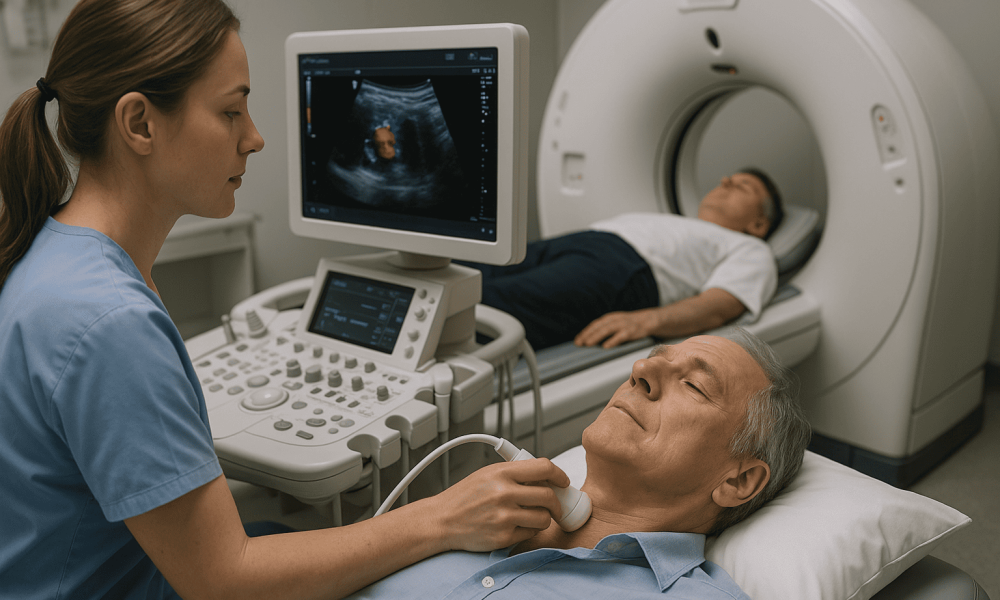
- Scanning: A handheld device called a transducer is moved over the neck to capture images of the carotid arteries. The transducer emits high-frequency sound waves that bounce off blood cells and arterial walls, creating real-time images.
- Imaging: The images are displayed on a monitor, allowing the technician or physician to assess the presence and extent of any arterial narrowing or plaque buildup.
- Duration: The entire procedure typically takes about 30 minutes and is painless, with no known risks associated.
Benefits of Carotid Ultrasound
Carotid ultrasound offers several advantages as a diagnostic tool:
- Non-Invasive: Does not require incisions or injections, making it a safe option for most patients.
- Real-Time Imaging: Provides immediate visualization of blood flow and arterial structures.
- No Radiation Exposure: Unlike CT scans, ultrasound does not use ionizing radiation, reducing potential risks.
- Cost-Effective: Generally less expensive compared to other imaging modalities like MRI or CT angiography.
- Portable: Can be performed in various settings, including bedside and outpatient clinics.
Limitations of Carotid Ultrasound
Despite its benefits, carotid ultrasound has certain limitations:
- Operator Dependent: The quality of the results can vary based on the technician’s expertise.
- Limited Penetration: May not effectively image deeper arterial segments or very small vessels.
- Inconclusive in Certain Cases: In cases of severe arterial calcification, ultrasound may have difficulty accurately assessing plaque characteristics.
Computed Tomography (CT) Scans: An Overview
Computed tomography (CT) scans are advanced imaging techniques that provide detailed cross-sectional images of the body. In the context of cardiovascular health, CT scans are invaluable for visualizing the arteries, detecting blockages, and planning surgical interventions.
Purpose of CT Scans in Cardiovascular Health
CT scans serve multiple purposes in cardiovascular diagnostics, including:
- Identifying Atherosclerosis: Detecting plaque buildup in coronary and other arteries.
- Assessing Arterial Anatomy: Providing detailed images of arterial structures for surgical planning.
- Evaluating Coronary Arteries: Performing coronary CT angiography to visualize coronary artery disease.
- Detecting Aneurysms: Identifying abnormal bulges in arterial walls that could lead to rupture.
Procedure of CT Scans
The CT scan procedure involves several key steps:
- Preparation: Patients may be required to fast for a few hours before the scan. Loose clothing and removal of metal objects are necessary to prevent interference with imaging.
- Contrast Material: A contrast dye, often containing iodine, may be injected intravenously to enhance the visibility of blood vessels and tissues.
- Positioning: The patient lies on a motorized table that slides into the CT scanner, a large, doughnut-shaped machine.
- Scanning: The scanner rotates around the patient, capturing multiple X-ray images from different angles. Advanced software reconstructs these images into detailed cross-sectional views.
- Duration: The procedure typically takes about 30 minutes, depending on the area being examined and the complexity of the case.
Benefits of CT Scans
CT scans offer several advantages in diagnosing and managing cardiovascular diseases:
- High Resolution: Provides detailed images of blood vessels, arteries, and other internal structures.
- Rapid Acquisition: Quickly captures comprehensive data, making it useful in emergency settings.
- 3D Imaging: Enables three-dimensional reconstruction of arterial anatomy, aiding in surgical planning and intervention.
- Versatility: Can be used to assess various cardiovascular conditions, from coronary artery disease to aneurysms.
Limitations of CT Scans
While CT scans are highly effective, they come with certain limitations:
- Radiation Exposure: Involves exposure to ionizing radiation, which carries a risk of cancer with cumulative exposure.
- Contrast Allergies: Some patients may experience allergic reactions to contrast dyes used during the scan.
- Cost: CT scans are generally more expensive than ultrasound and other imaging modalities.
- Limited Soft Tissue Contrast: While excellent for visualizing bones and vessels, CT scans are less effective for soft tissue differentiation compared to MRI.
Comparing Carotid Ultrasound and CT Scans
Both carotid ultrasound and CT scans are essential tools in cardiovascular diagnostics, each with its unique advantages and applications. Understanding their differences helps in selecting the appropriate test based on the clinical scenario.
Carotid Ultrasound vs. CT Angiography
- Invasiveness: Carotid ultrasound is non-invasive and does not require contrast, whereas CT angiography involves contrast injection.
- Radiation: Carotid ultrasound does not use ionizing radiation, making it safer for repeated use, while CT scans expose patients to radiation.
- Detail and Resolution: CT scans provide higher resolution images and can visualize deeper arterial structures, while ultrasound is limited to surface and medium-depth vessels.
- Cost and Accessibility: Ultrasound is generally more affordable and widely accessible compared to CT scans, which are costlier and require specialized equipment.
- Use Cases: Ultrasound is ideal for initial screening and monitoring of carotid artery disease, whereas CT scans are preferred for detailed anatomical assessment and surgical planning.
When to Use Each Diagnostic Test
Choosing between carotid ultrasound and CT scans depends on various factors, including the patient’s symptoms, risk factors, and the specific clinical question being addressed.
- Carotid Ultrasound: Best suited for routine screening of carotid artery disease, evaluating blood flow dynamics, and monitoring known arterial stenosis. It is particularly useful in patients with a history of stroke, transient ischemic attacks (TIAs), or significant risk factors like hypertension and smoking.
- CT Scans: Indicated for comprehensive assessment of arterial anatomy, planning surgical interventions, evaluating complex or multi-vessel disease, and detecting arterial anomalies or aneurysms. CT angiography is especially valuable in acute settings, such as suspected pulmonary embolism or in pre-operative planning for vascular surgery.
Roles in Disease Management
Both carotid ultrasound and CT scans play pivotal roles in the diagnosis, treatment planning, and monitoring of cardiovascular diseases.
Carotid Ultrasound in Disease Management
- Diagnosis: Identifies the presence and extent of carotid artery stenosis.
- Treatment Planning: Determines the need for medical therapy, lifestyle modifications, or surgical interventions like carotid endarterectomy.
- Monitoring: Tracks the progression of arterial disease and the effectiveness of treatments over time.
CT Scans in Disease Management
- Surgical Planning: Provides detailed images for planning coronary artery bypass grafting (CABG) or stent placement.
- Risk Assessment: Evaluates the severity of arterial blockages and the overall burden of atherosclerosis.
- Post-Treatment Evaluation: Assesses the success of interventions and detects any complications or residual disease.
Preparing for Carotid Ultrasound and CT Scans
Proper preparation ensures the accuracy of diagnostic tests and enhances patient comfort during the procedures.
Preparing for Carotid Ultrasound
- Clothing: Wear loose-fitting clothing that allows easy access to the neck area.
- Hydration: Staying well-hydrated can improve the quality of ultrasound images.
- Avoiding Topical Products: Remove any lotions, oils, or hair products from the neck to ensure clear contact with the ultrasound gel.
- Fasting: Generally, no fasting is required unless specified by the healthcare provider.
Preparing for CT Scans
- Fasting: Patients may be required to fast for a few hours before the scan, especially if contrast dye is used.
- Contrast Allergy: Inform the healthcare provider of any known allergies to contrast materials or iodine.
- Medication Management: Some medications may need to be adjusted or paused before the scan. Consult with the healthcare provider.
- Clothing: Wear comfortable, metal-free clothing and remove any jewelry or metal accessories.
- Hydration: Drinking plenty of water before the scan can aid in the elimination of contrast dye after the procedure.
Post-Procedure Considerations
After undergoing carotid ultrasound or CT scans, certain post-procedure steps ensure patient safety and optimal outcomes.
After Carotid Ultrasound
- No Recovery Time: Patients can resume normal activities immediately after the procedure.
- Follow-Up: Discuss the results with the healthcare provider to determine the next steps in management.
After CT Scans
- Hydration: Increase fluid intake to help flush the contrast dye from the body.
- Monitoring: Observe for any adverse reactions to the contrast material, such as rash, itching, or difficulty breathing, and seek immediate medical attention if they occur.
- Activity: Most patients can resume normal activities, but specific instructions may vary based on the individual’s health status and the reason for the scan.
For an overview of heart and vascular wellness—including atherosclerosis prevention basics—see: Cardiovascular Health: The Definitive Guide to Heart and Vascular Wellness.
Conclusion
Carotid ultrasound and CT scans are indispensable tools in the realm of cardiovascular diagnostics. Carotid ultrasound offers a non-invasive, cost-effective method for assessing carotid artery health and preventing strokes, while CT scans provide detailed anatomical insights essential for comprehensive cardiovascular evaluation and surgical planning. Understanding the procedures, benefits, and limitations of these diagnostic tests empowers patients and healthcare providers to make informed decisions, ensuring timely and effective management of cardiovascular diseases. By leveraging these advanced imaging techniques, the healthcare community can continue to enhance diagnostic accuracy, optimize treatment strategies, and ultimately improve patient outcomes in the fight against cardiovascular ailments.
