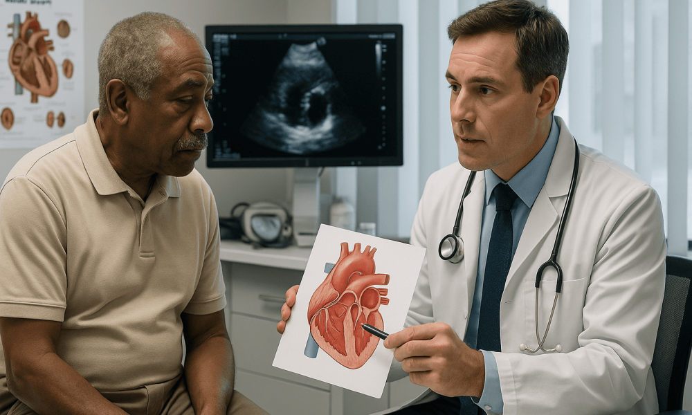
Aortic Stenosis: Causes and Treatments
Aortic stenosis (AS) is a prevalent and serious heart valve disease characterized by the narrowing of the aortic valve opening, which restricts blood flow from the left ventricle into the aorta and onward to the rest of the body. This condition forces the heart to work harder to pump blood, leading to various symptoms and increasing the risk of severe complications such as heart failure and sudden cardiac death. Understanding the causes and available treatments for aortic stenosis is crucial for effective management and improved patient outcomes.
To put valve conditions into the wider framework of heart and vascular wellness, see: Cardiovascular Health: The Definitive Guide to Heart and Vascular Wellness.
Understanding Aortic Stenosis
Aortic stenosis is one of the most common valvular heart diseases, especially in older adults. It involves the progressive calcification and thickening of the aortic valve leaflets, which impedes the valve’s ability to open fully.
Anatomy of the Aortic Valve
The aortic valve is located between the left ventricle of the heart and the aorta. It typically has three leaflets (tricuspid), although some individuals may have a bicuspid aortic valve, which is a congenital variation with two leaflets. The primary function of the aortic valve is to ensure unidirectional blood flow from the heart into the aorta during ventricular contraction (systole) and to prevent backflow during ventricular relaxation (diastole).
Pathophysiology of Aortic Stenosis
The narrowing of the aortic valve leads to increased resistance against which the left ventricle must pump, resulting in left ventricular hypertrophy (thickening of the heart muscle). Over time, the heart may become less efficient, leading to symptoms such as chest pain, shortness of breath, and syncope. Severe aortic stenosis can significantly impair the heart’s ability to supply adequate blood to the body.
Causes of Aortic Stenosis
Aortic stenosis can result from several underlying causes, each contributing to the progressive narrowing of the aortic valve.
Age-Related Calcific Degeneration
The most common cause of aortic stenosis in adults over the age of 65 is degenerative calcific aortic stenosis. This process involves the gradual accumulation of calcium deposits on the aortic valve leaflets, leading to their stiffening and fusion. Over time, this calcification reduces the mobility of the valve, limiting its opening.
Congenital Bicuspid Aortic Valve
Approximately 1-2% of the population is born with a bicuspid aortic valve, where the valve has two leaflets instead of the usual three. This congenital anomaly predisposes individuals to earlier development of aortic stenosis and other valve-related issues compared to those with a tricuspid valve.
Rheumatic Fever
Rheumatic fever, a complication of untreated or inadequately treated streptococcal throat infections, can cause inflammatory damage to the heart valves, including the aortic valve. Although less common in developed countries due to improved antibiotic use, rheumatic aortic stenosis remains a significant cause in developing regions.
Radiation Therapy
Individuals who have undergone radiation therapy to the chest area, particularly for cancers like Hodgkin’s lymphoma or breast cancer, may develop aortic stenosis years later. Radiation can cause fibrosis and calcification of the heart valves, including the aortic valve.
Other Causes
Less common causes of aortic stenosis include infective endocarditis (infection of the heart valves), certain metabolic disorders, and connective tissue diseases that affect the heart valves’ structure and function.
Symptoms of Aortic Stenosis
Aortic stenosis may remain asymptomatic for years as the condition progresses slowly. However, as the valve narrowing becomes more severe, symptoms begin to manifest, signaling the need for medical intervention.
Common Symptoms
- Chest Pain (Angina): A feeling of tightness, pressure, or pain in the chest, especially during physical exertion.
- Shortness of Breath (Dyspnea): Difficulty breathing during activities or even at rest in severe cases.
- Fainting (Syncope): Sudden loss of consciousness, often triggered by exertion or emotional stress.
- Fatigue: Persistent tiredness and reduced exercise capacity due to decreased cardiac output.
Less Common Symptoms
- Heart Palpitations: Awareness of irregular or rapid heartbeats.
- Swelling (Edema): Fluid retention in the legs, ankles, or abdomen.
- Reduced Appetite and Weight Loss: Due to decreased blood flow and energy levels.
Diagnosis of Aortic Stenosis
Early diagnosis of aortic stenosis is essential for timely management and prevention of complications. The diagnostic process typically involves a combination of clinical evaluation and imaging studies.
Physical Examination
During a physical exam, a healthcare provider may detect a heart murmur, which is a sound produced by turbulent blood flow through the narrowed aortic valve. The murmur of aortic stenosis is typically a systolic ejection murmur best heard at the right upper sternal border and may radiate to the carotid arteries.
Echocardiography
Echocardiography is the primary diagnostic tool for aortic stenosis. It uses ultrasound waves to create detailed images of the heart valves and chambers, allowing for assessment of valve structure, function, and the degree of stenosis.
- Transthoracic Echocardiogram (TTE): A non-invasive procedure where the ultrasound probe is placed on the chest to visualize the heart valves and measure the pressure gradients across the aortic valve.
- Transesophageal Echocardiogram (TEE): Involves inserting the probe into the esophagus for clearer images, especially useful in complex cases or when TTE results are inconclusive.
Cardiac Catheterization and Coronary Angiography
These invasive procedures involve threading a catheter into the heart to measure pressures within the chambers and assess the severity of aortic stenosis. Coronary angiography may also be performed to evaluate for concurrent coronary artery disease, which is common in patients with aortic stenosis.
Cardiac MRI and CT Scans
Advanced imaging techniques like cardiac MRI and CT scans provide additional information about the heart’s structure and function. They can help in assessing the extent of valve calcification and planning for surgical interventions.
Electrocardiogram (ECG)
An ECG records the heart’s electrical activity and can detect signs of left ventricular hypertrophy, arrhythmias, or previous myocardial infarctions, which may coexist with aortic stenosis.
Chest X-Ray
A chest X-ray can reveal an enlarged heart silhouette, calcification of the aortic valve, and signs of pulmonary congestion or edema.
Treatment Options for Aortic Stenosis
The management of aortic stenosis depends on the severity of the condition and the presence of symptoms. Treatment options range from medical management to surgical and interventional procedures.
Medical Management
In the early stages of aortic stenosis, when symptoms are mild or absent, medical management focuses on controlling risk factors and preventing disease progression.
- Medications: Diuretics may be prescribed to manage fluid retention and reduce symptoms of heart failure. Beta-blockers and ACE inhibitors can help manage hypertension and reduce cardiac workload.
- Lifestyle Modifications: Adopting a heart-healthy diet, engaging in regular physical activity, quitting smoking, and limiting alcohol intake are essential for overall cardiovascular health.
However, it is important to note that medical management alone cannot reverse aortic stenosis. When the condition progresses or symptoms worsen, more definitive treatments are necessary.
Surgical Treatments
Surgical intervention is the most effective treatment for severe aortic stenosis. The primary surgical options include aortic valve replacement and, in some cases, aortic valve repair.
Aortic Valve Replacement (AVR)
AVR involves removing the diseased aortic valve and replacing it with a prosthetic valve. There are two main types of prosthetic valves:
- Mechanical Valves: Made from durable materials like titanium or carbon, these valves are long-lasting but require lifelong anticoagulation therapy to prevent blood clots.
- Bioprosthetic (Tissue) Valves: Made from animal tissues (usually pig or cow), these valves typically do not require long-term anticoagulation but may have a shorter lifespan compared to mechanical valves.
Benefits: AVR effectively relieves symptoms, improves heart function, and enhances quality of life. It also reduces the risk of complications such as heart failure and sudden cardiac death.
Risks: As with any major surgery, risks include infection, bleeding, stroke, and complications related to anesthesia.
Aortic Valve Repair
In select cases, particularly when the valve leaflets are sufficiently intact, surgical repair may be possible. Valve repair techniques include reshaping, relining, or reconstructing the valve to restore its function.
Benefits: Preserving the patient’s native valve reduces the need for anticoagulation therapy and can lead to better hemodynamic performance.
Limitations: Valve repair is not suitable for all cases, especially when there is significant calcification or structural damage to the valve.
Interventional Procedures
For patients who are at high risk for traditional open-heart surgery, less invasive interventional procedures offer effective alternatives.
Transcatheter Aortic Valve Replacement (TAVR)
TAVR is a minimally invasive procedure where a prosthetic valve is delivered via a catheter, usually through the femoral artery, and implanted within the existing aortic valve. This procedure is particularly beneficial for elderly patients or those with significant comorbidities that make surgery risky.
- Benefits: Shorter recovery time, reduced procedural risks, and the ability to perform the procedure without open-heart surgery.
- Risks: Potential complications include vascular complications, valve leakage, and the need for a permanent pacemaker.
TAVR has become increasingly popular due to its effectiveness and improved safety profile, making it a viable option for a broader range of patients.
Post-Treatment Care and Management
Following treatment for aortic stenosis, ongoing care is essential to ensure the success of the intervention and to maintain optimal heart health.
Regular Follow-ups
Patients require routine check-ups with their cardiologist to monitor heart function, assess the performance of the prosthetic valve, and detect any potential complications early.
Medication Adherence
Adhering to prescribed medications, such as anticoagulants for patients with mechanical valves, is crucial for preventing complications like blood clots and ensuring the longevity of the treatment outcomes.
Lifestyle Modifications
Continuing with a heart-healthy lifestyle is vital for maintaining heart health and preventing the progression of other cardiovascular conditions. This includes:
- Balanced Diet: Emphasizing fruits, vegetables, whole grains, lean proteins, and low-fat dairy products while limiting saturated fats, trans fats, cholesterol, and sodium.
- Regular Exercise: Engaging in moderate physical activity, as recommended by a healthcare provider, to strengthen the heart and improve overall cardiovascular fitness.
- Smoking Cessation: Avoiding tobacco use significantly reduces the risk of further heart disease and improves overall health outcomes.
- Weight Management: Maintaining a healthy weight helps reduce the burden on the heart and lowers the risk of hypertension and diabetes.
- Stress Management: Implementing stress-reduction techniques such as meditation, yoga, or deep-breathing exercises can benefit heart health.
Prognosis and Outcomes
The prognosis for patients with aortic stenosis has improved significantly with advancements in diagnostic and treatment modalities. Early detection and timely intervention are key factors in enhancing survival rates and quality of life. Surgical and interventional treatments have demonstrated substantial benefits in symptom relief, functional improvement, and reduction in mortality rates.
Factors Influencing Prognosis
Several factors can influence the prognosis of aortic stenosis, including:
- Severity of Stenosis: Severe aortic stenosis has a more guarded prognosis compared to mild or moderate cases.
- Presence of Symptoms: Symptomatic aortic stenosis requires prompt intervention to prevent adverse outcomes.
- Age and Overall Health: Younger patients and those in good overall health typically have better outcomes following treatment.
- Timing of Intervention: Early surgical or interventional treatment before the onset of severe symptoms leads to improved survival rates.
- Associated Medical Conditions: The presence of other cardiovascular or systemic conditions can impact the overall prognosis.
Long-term Outcomes
With appropriate treatment, many patients experience significant improvements in symptoms and quality of life. Mechanical and bioprosthetic valves generally function well, although regular monitoring is necessary to detect and address any issues that may arise over time. Advances in TAVR technology continue to expand the treatment options, making valve replacement accessible to a wider patient population with favorable long-term outcomes.
Prevention of Aortic Stenosis
While some causes of aortic stenosis, such as congenital defects, cannot be prevented, certain measures can help reduce the risk or slow the progression of the condition.
- Managing Risk Factors: Controlling hypertension, diabetes, and cholesterol levels through lifestyle changes and medications can help prevent the development of aortic stenosis.
- Avoiding Infections: Prompt treatment of infections, particularly rheumatic fever, can prevent valve damage and subsequent stenosis.
- Healthy Lifestyle: Maintaining a balanced diet, engaging in regular physical activity, avoiding smoking, and limiting alcohol intake contribute to overall heart health and reduce the risk of valvular diseases.
- Regular Medical Check-ups: Routine health evaluations can aid in the early detection and management of heart conditions that may lead to aortic stenosis.
For the big-picture basics of cardiovascular health (including risk factors and prevention), read the cornerstone guide: Cardiovascular Health: The Definitive Guide to Heart and Vascular Wellness.
Conclusion
Aortic stenosis is a significant cardiovascular condition that requires timely diagnosis and effective management to prevent severe complications and improve patient outcomes. Understanding the causes, recognizing the symptoms, and seeking appropriate medical care are essential steps in addressing this condition. Advances in surgical and interventional treatments, particularly the development of minimally invasive procedures like TAVR, have revolutionized the management of aortic stenosis, offering better outcomes and enhanced quality of life for patients. Additionally, adopting a heart-healthy lifestyle and managing underlying risk factors play a crucial role in preventing the onset and progression of aortic stenosis. Ongoing research and technological innovations continue to improve the prognosis for individuals affected by this condition, underscoring the importance of continued advancements in cardiovascular medicine.
