Different Types of Mammograms: 2D vs. 3D Screening
Mammography is a critical tool in the early detection of breast cancer, offering a window into your breast health with remarkable clarity. In recent years, two main types of mammograms have emerged: the traditional 2D mammogram and the advanced 3D mammogram, also known as digital breast tomosynthesis. While both techniques aim to spot early signs of cancer, they work in slightly different ways and offer unique benefits. If you’ve ever wondered what sets them apart or which one might be best for you, you’re in the right place. Let’s explore these screening methods together in a friendly, accessible manner.
For the essentials of preventive testing, screening schedules, and early detection, see our cornerstone guide: Health Screenings: Your Guide to Preventive Testing and Early Detection.
Understanding Mammography
Mammography is essentially an X-ray of the breast, designed to detect abnormal growths or changes in breast tissue. The idea behind any mammogram is to capture detailed images that can reveal hidden abnormalities long before they become palpable lumps. This early detection is key in improving treatment outcomes and reducing the risk of advanced disease.
Imagine your breast as a complex landscape, with different layers and contours. Traditional 2D mammography captures this landscape in a single, flat image. On the other hand, 3D mammography takes multiple images from various angles, which are then compiled into a three-dimensional view of your breast tissue. This difference in technique has sparked much discussion among healthcare providers and patients alike.
2D Mammography: The Classic Approach
For decades, 2D mammography has been the standard method of screening for breast cancer. With 2D mammograms, your breast is compressed between two plates while an X-ray machine takes a single, flat image. This method has proven effective over many years, helping countless women with early detection.
There are several advantages to 2D mammography:
- Time-Tested Reliability: 2D mammograms have been used successfully for decades, providing a wealth of clinical data that supports their use.
- Quick Procedure: The imaging process is relatively fast, which can help reduce anxiety for many patients.
- Widespread Availability: Because of its long history, most imaging centers and hospitals offer 2D mammography, making it easily accessible to many women.
However, 2D mammography does have its limitations. The primary challenge lies in its ability to capture overlapping tissue. In women with dense breast tissue, for example, the superimposition of tissues can sometimes mask tumors, potentially leading to false negatives or the need for additional imaging tests.
3D Mammography: A Modern Innovation
Enter 3D mammography, an innovation that builds upon the foundation of 2D imaging. Also known as digital breast tomosynthesis, 3D mammography takes a series of X-ray images of the breast from multiple angles. These images are then reconstructed into a three-dimensional picture, allowing radiologists to examine breast tissue layer by layer.
Some of the standout benefits of 3D mammography include:
- Improved Detection Rates: The layered images reduce the chance of tissue overlap, which can make it easier to spot small cancers hidden in dense breast tissue.
- Fewer Callbacks: Because the 3D images provide more detailed views, there is often less need for additional follow-up tests after the initial screening.
- Enhanced Clarity: The tomosynthesis technique allows for a clearer differentiation between benign and suspicious areas, aiding in more accurate diagnoses.
While 3D mammography is a promising technology, it is not without challenges. For instance, it may take slightly longer to perform compared to the traditional 2D exam. In addition, not every healthcare facility has the latest 3D equipment, which might limit its accessibility in some regions.
Comparing 2D and 3D Mammograms
At first glance, the difference between 2D and 3D mammography might seem subtle, but the implications are significant. Both methods use the same underlying technology—X-rays—to capture images of the breast. However, the way these images are processed and interpreted sets them apart.
Here are some key points to consider when comparing the two techniques:
- Image Depth: 2D mammograms provide a single flat image, while 3D mammograms offer depth by displaying multiple slices of the breast tissue. This extra dimension can be especially beneficial for women with dense breasts.
- Detection Accuracy: Studies have shown that 3D mammography can increase the detection rate of breast cancer and reduce the number of false positives, leading to fewer unnecessary callbacks and biopsies.
- Procedure Duration: A 2D mammogram is typically quicker, but the slight increase in time for a 3D exam is often offset by the enhanced diagnostic confidence it provides.
- Radiation Exposure: Although 3D mammography uses a bit more radiation than 2D, the levels remain within safe limits. Advances in technology continue to minimize exposure while maximizing image quality.
It’s important to note that the choice between 2D and 3D mammography isn’t about one being inherently superior to the other. Rather, it’s about what works best for your individual needs and risk factors. Many healthcare providers now offer both options, and in some cases, a combination of the two is used to achieve the most comprehensive screening.
What to Expect During a Mammogram
Whether you’re scheduled for a 2D or 3D mammogram, the procedure itself follows a similar general process. Here’s a brief overview to help you feel more comfortable about what lies ahead:
Before the Exam
Your appointment will usually begin with a brief consultation where you can discuss your medical history and any concerns you might have. The technologist will explain the process and answer questions to help ease any anxiety.
It’s a good idea to avoid using deodorants, powders, or lotions on your breasts and underarms on the day of your exam, as these substances can interfere with the quality of the images.
During the Exam
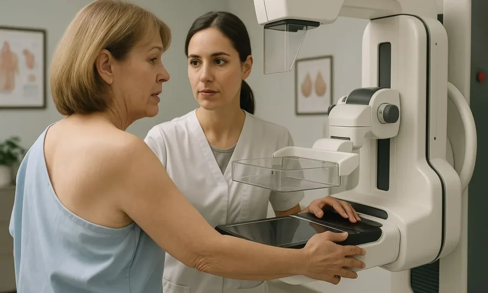
For the actual mammogram, you will stand in front of the imaging machine. Your breast is gently placed on a flat surface, and a compression paddle is used to compress the tissue. Although this compression might feel uncomfortable, it is essential for spreading out the breast tissue and capturing clear images.
If you’re undergoing a 3D mammogram, the process involves taking several images from different angles. The machine will rotate slightly as it captures these slices, which are later compiled into a detailed, layered image of your breast.
The entire procedure typically takes only about 15 to 30 minutes. While the compression may be momentarily uncomfortable, the quick process helps to minimize any prolonged discomfort.
After the Exam
Once the imaging is complete, you can dress and resume your normal activities immediately. Your radiologist will review the images and send a report to your doctor, who will then discuss the results with you. It’s common to feel a mix of relief and curiosity after the exam, knowing that you have taken an important step in monitoring your breast health.
Making an Informed Decision
Choosing between 2D and 3D mammography ultimately depends on your personal health profile, breast density, and the recommendations of your healthcare provider. Both techniques offer valuable insights, and the decision should be tailored to your individual circumstances.
If you have a family history of breast cancer or denser breast tissue, you might benefit from the enhanced detection capabilities of 3D mammography. Conversely, if you are at average risk and prefer a quicker procedure, a 2D mammogram may be sufficient.
Discuss your options with your doctor. They can explain how each method aligns with your specific health needs and help you choose the best screening strategy. Remember, the goal is early detection and peace of mind, and both methods are effective tools in achieving that goal.
Looking Ahead: The Future of Mammography
Advances in medical imaging continue to refine and enhance mammography techniques. Research is ongoing to further reduce radiation exposure, improve image clarity, and tailor screening protocols to individual risk factors. As technology evolves, you can expect even more personalized and accurate screenings, ultimately contributing to better outcomes and improved breast health for women everywhere.
The journey toward better breast health is ever-evolving, and staying informed about your options is a key part of that process. By understanding the differences between 2D and 3D mammography, you are better equipped to take charge of your health and make decisions that align with your personal needs.
To place mammograms within a complete prevention and screening plan, start here: the Health Screenings cornerstone guide.
Final Thoughts: Empowerment Through Knowledge
In the realm of breast health, knowledge is truly empowering. Whether you opt for a 2D or 3D mammogram, each method plays an important role in the early detection of breast cancer. By understanding how these techniques work, their benefits, and what to expect during your screening, you can approach your next exam with confidence and assurance.
Remember, regular screening is one of the most effective ways to protect your health. Take the time to discuss your options with your healthcare provider, ask questions, and stay proactive about your well-being. With every informed decision, you’re not only taking care of your health today—you’re investing in a brighter, healthier future.
So, as you prepare for your next mammogram, embrace the process with the knowledge that you are in control. Whether you choose the classic approach of 2D imaging or the enhanced detail of 3D screening, every step you take is a step toward early detection, effective treatment, and lasting peace of mind.
